alb10694504
MRI Pilocytic Astrocytoma
| Partager |
|---|
 Pinterest Pinterest |
 Twitter Twitter |
 Facebook Facebook |
 Copier le lien Copier le lien |
 Email Email |
|
Ajouter à une autre Lightbox |
|
Ajouter à une autre Lightbox |


Avez-vous déjà un compte? S'identifier
Vous n'avez pas de compte ? S'inscrire
Acheter cette image

Titre:
MRI Pilocytic Astrocytoma
Légende:
Voir la traduction automatique
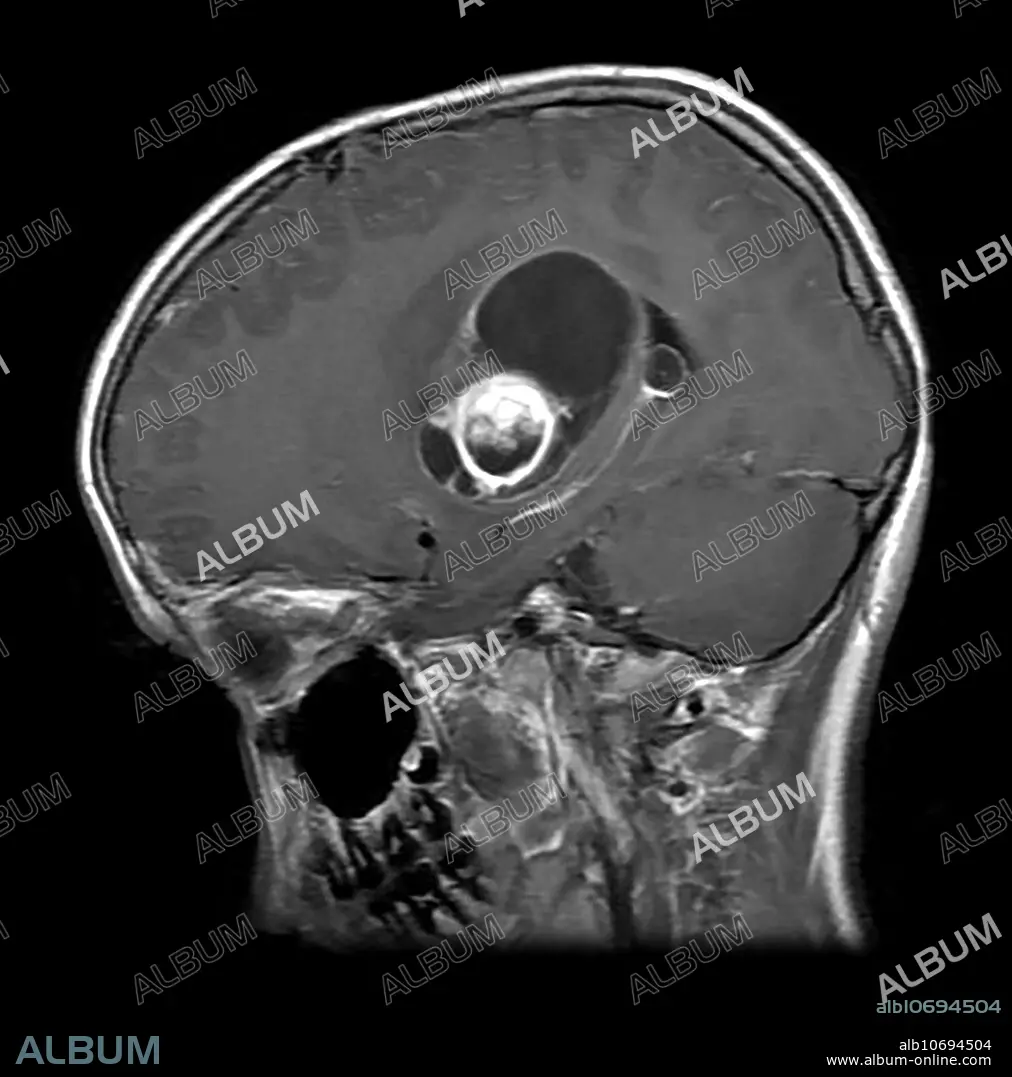
This axial (cross sectional) T1 weighted MR image with contrast shows a partially cystic and solid enhancing mass in the basal ganglia, internal capsule and thalamic regions with associate mass effect in a 25 year old. This represents a WHO grade 1 astrocytoma called a pilocytic astrocytoma.
Personnalités:
Crédit:
Album / Living Art Enterprises, LLC/Science Source
Autorisations:
Modèle: Non - Propriété: Non
Questions sur les droits?
Questions sur les droits?
Taille de l'image:
3900 x 3947 px | 44.0 MB
Taille d'impression:
33.0 x 33.4 cm | 13.0 x 13.2 in (300 dpi)
Mots clés:
