alb10618863
Microvilli in Intestinal Epithelium, TEM
| Partager |
|---|
 Pinterest Pinterest |
 Twitter Twitter |
 Facebook Facebook |
 Copier le lien Copier le lien |
 Email Email |
|
Ajouter à une autre Lightbox |
|
Ajouter à une autre Lightbox |


Avez-vous déjà un compte? S'identifier
Vous n'avez pas de compte ? S'inscrire
Acheter cette image.
Sélectionnez l'usage:

Titre:
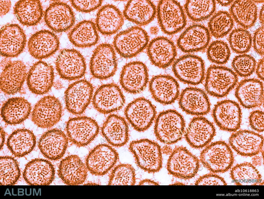
Microvilli in Intestinal Epithelium, TEM
Légende:
Voir la traduction automatique
Colour enhanced transmission electron micrograph of microvilli of the brush border of cat intestinal epithelium. This transverse section of intestinal brush border shows the actin filaments that form the core of each microvillus. At the villus tip, the relation of the filaments to the membrane is obscured by a layer of dense amorphous material associated with the inner aspect of the membrane.
Crédit:
Album / Science Source / Don W. Fawcett
Autorisations:
Taille de l'image:
4437 x 3119 px | 39.6 MB
Taille d'impression:
37.6 x 26.4 cm | 14.8 x 10.4 in (300 dpi)
Mots clés:
