alb10608671
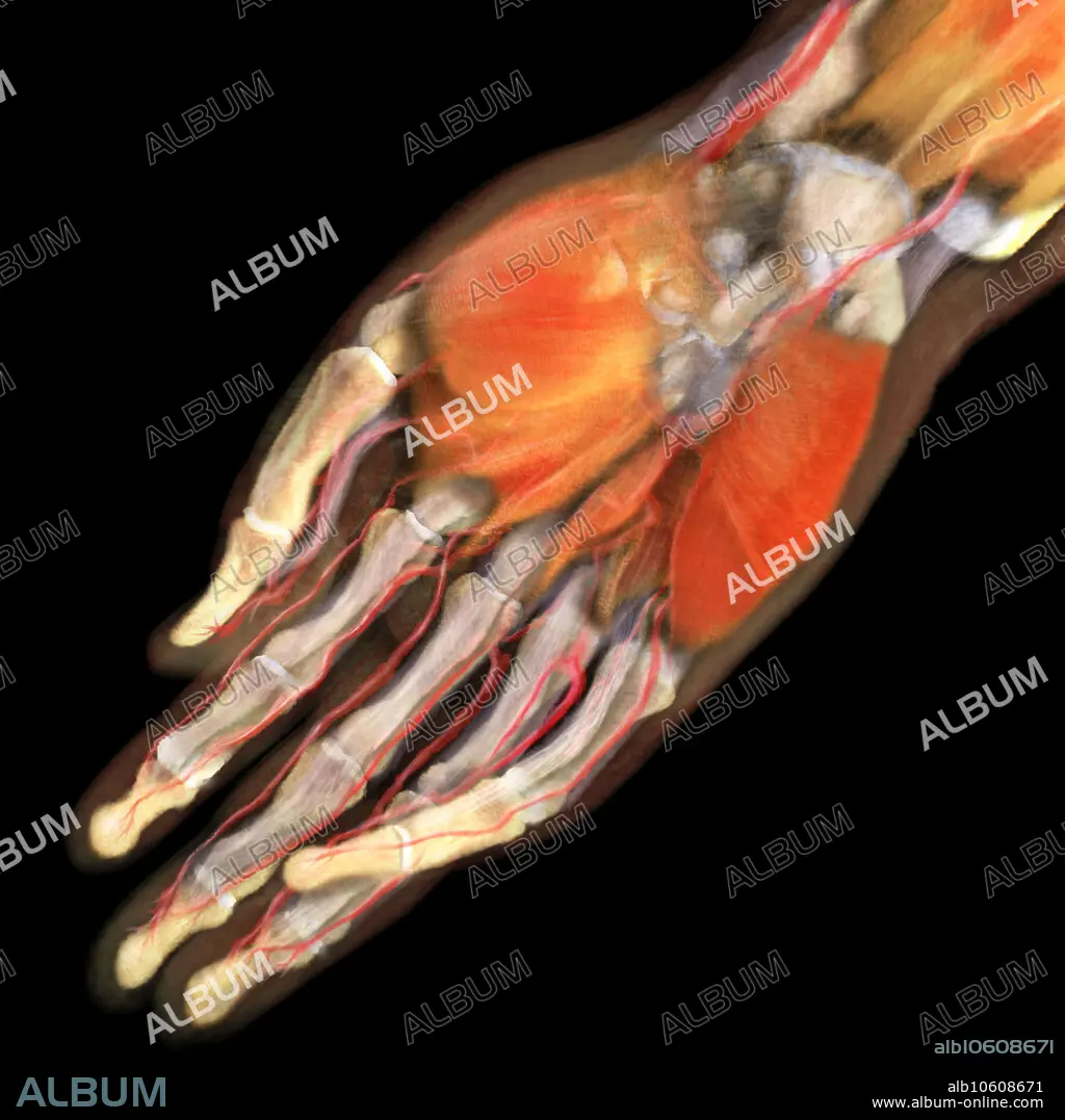
Right Hand Anatomy, Ventral View
| Partager |
|---|
 Pinterest Pinterest |
 Twitter Twitter |
 Facebook Facebook |
 Copier le lien Copier le lien |
 Email Email |
|
Ajouter à une autre Lightbox |
|
Ajouter à une autre Lightbox |


Avez-vous déjà un compte? S'identifier
Vous n'avez pas de compte ? S'inscrire
Acheter cette image.
Sélectionnez l'usage:

Titre:
Right Hand Anatomy, Ventral View
Légende:
Voir la traduction automatique
3D visualization based on scanned human data of the ventral side of a human right hand. On the palmar side, the thenar and hypothenar muscles are visible; lying beneath them are the metacarpal bones. The phalanges are revealed with the palmar digital arteries running also aside them.
Crédit:
Album / Science Source / ANATOMICAL TRAVELOGUE
Autorisations:
Modèle: Non - Propriété: Non
Questions sur les droits?
Questions sur les droits?
Taille de l'image:
2000 x 2000 px | 11.4 MB
Taille d'impression:
16.9 x 16.9 cm | 6.7 x 6.7 in (300 dpi)
