alb3886157
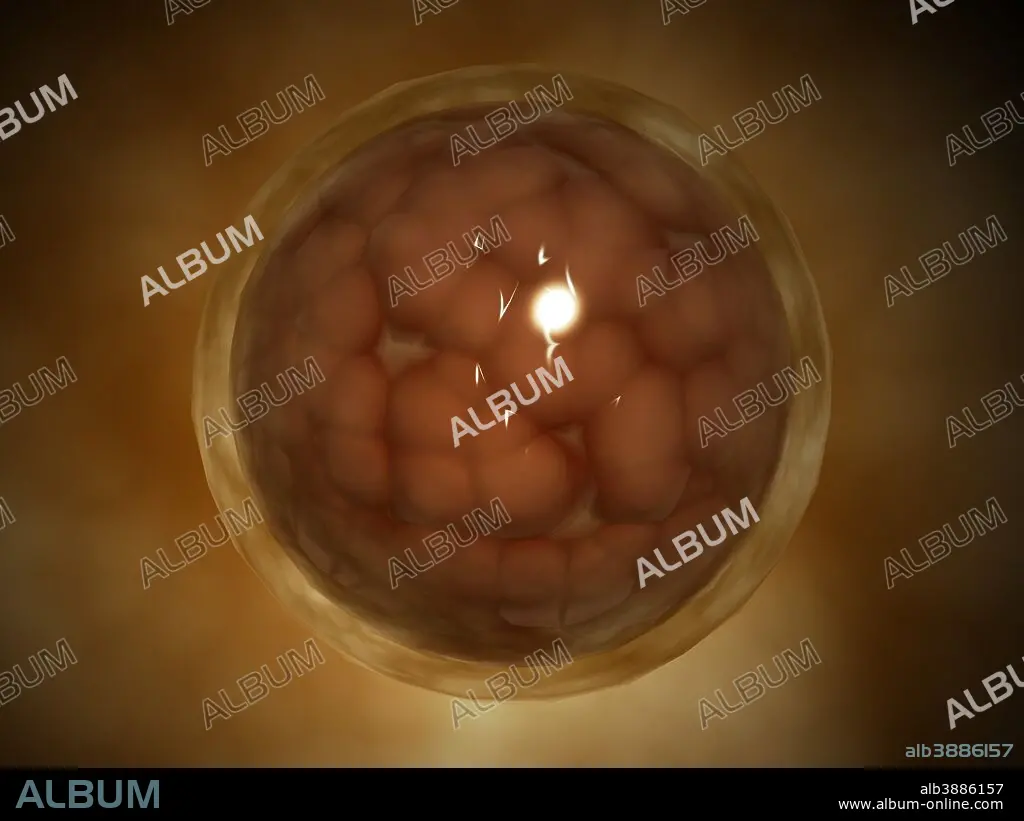
Microscopic view of a blastula during pregnancy.
| Partager |
|---|
 Pinterest Pinterest |
 Twitter Twitter |
 Facebook Facebook |
 Copier le lien Copier le lien |
 Email Email |
|
Ajouter à une autre Lightbox |
|
Ajouter à une autre Lightbox |


Avez-vous déjà un compte? S'identifier
Vous n'avez pas de compte ? S'inscrire
Acheter cette image

Titre:
Microscopic view of a blastula during pregnancy.
Légende:
Voir la traduction automatique
Microscopic view of a blastula during pregnancy. After the cleavage has produced over 100 cells, the embryo is called a blastula. The blastula is usually a spherical layer of cells (the blastoderm) surrounding a fluid-filled or yolk-filled cavity (the blastocoel).
Crédit:
Album / Stocktrek Images
Autorisations:
Modèle: Non - Propriété: Non
Questions sur les droits?
Questions sur les droits?
Taille de l'image:
4920 x 3690 px | 51.9 MB
Taille d'impression:
41.7 x 31.2 cm | 16.4 x 12.3 in (300 dpi)
Mots clés:
