alb3788974
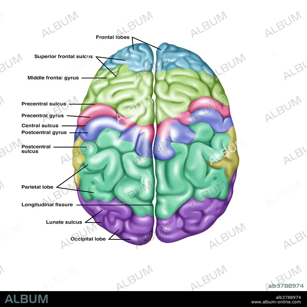
Top View of Normal Brain, Illustration
| Partager |
|---|
 Pinterest Pinterest |
 Twitter Twitter |
 Facebook Facebook |
 Copier le lien Copier le lien |
 Email Email |
|
Ajouter à une autre Lightbox |
|
Ajouter à une autre Lightbox |


Avez-vous déjà un compte? S'identifier
Vous n'avez pas de compte ? S'inscrire
Acheter cette image.
Sélectionnez l'usage:

Titre: Top View of Normal Brain, Illustration
Légende: Voir la traduction automatique
Illustration showing anatomy of a normal brain in a superior (top) view. Noted from top to bottom on the left side are the following: frontal lobes, superior frontal sulcus, middle frontal gyrus, precentral sulcus, precentral gyrus, central sulcus, postcentral gyrus, postcentral sulcus, parietal lobe, longitudinal fissure, lunate sulcus, and the occipital lobe.
Illustration showing anatomy of a normal brain in a superior (top) view. Noted from top to bottom on the left side are the following: frontal lobes, superior frontal sulcus, middle frontal gyrus, precentral sulcus, precentral gyrus, central sulcus, postcentral gyrus, postcentral sulcus, parietal lobe, longitudinal fissure, lunate sulcus, and the occipital lobe.
Crédit: Album / Science Source / Gwen Shockey
Autorisations: ? Autorisation de modèle: Non - ? Autorisation de propriété: Non
Questions sur les droits?
Questions sur les droits?
Taille de l'image: 4337 × 4110 px | 51.0 MB
Taille d'impression: 36.7 × 34.8 cm | 1707.5 × 1618.1 in (300 dpi)
Mots clés: ANATOMIE • GRAPHIQUE • ILLUSTRATION
