alb5575977
3D illustration of spherical-shaped, measles virus particle.
| Partager |
|---|
 Pinterest Pinterest |
 Twitter Twitter |
 Facebook Facebook |
 Copier le lien Copier le lien |
 Email Email |
|
Ajouter à une autre Lightbox |
|
Ajouter à une autre Lightbox |


Avez-vous déjà un compte? S'identifier
Vous n'avez pas de compte ? S'inscrire
Acheter cette image.
Sélectionnez l'usage:

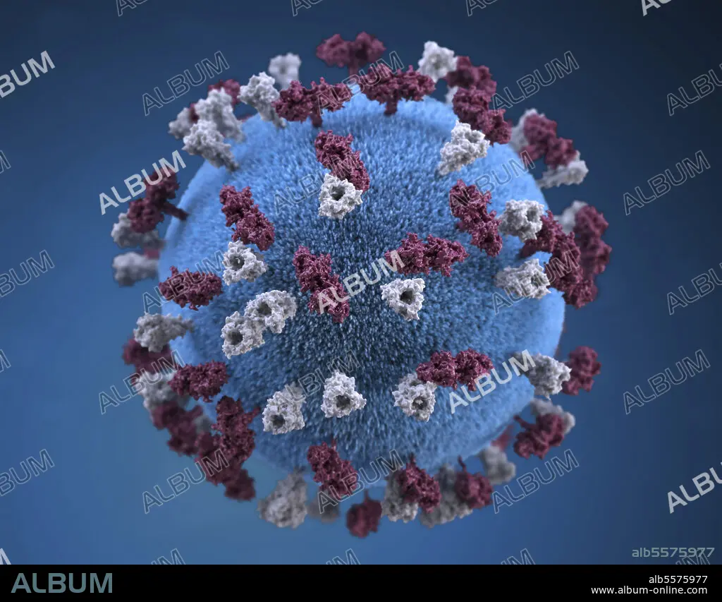
Titre: 3D illustration of spherical-shaped, measles virus particle.
Légende: Voir la traduction automatique
3D illustration of spherical-shaped, measles virus particle, that is studded with glycoprotein tubercles. These tubercular studs colorized maroon, are known as H-proteins (hemagglutinin), while those colorized gray, represented what are referred to as F-proteins (fusion). The F-protein is responsible for fusion of the virus and host cell membranes, viral penetration, and hemolysis. The H-protein is responsible for the binding of virions to cells. Both types of proteinaceous studs are embedded in the particle envelopes lipid bilayer.
3D illustration of spherical-shaped, measles virus particle, that is studded with glycoprotein tubercles. These tubercular studs colorized maroon, are known as H-proteins (hemagglutinin), while those colorized gray, represented what are referred to as F-proteins (fusion). The F-protein is responsible for fusion of the virus and host cell membranes, viral penetration, and hemolysis. The H-protein is responsible for the binding of virions to cells. Both types of proteinaceous studs are embedded in the particle envelopes lipid bilayer.
Crédit: Album / Stocktrek Images
Taille de l'image: 3600 × 2813 px | 29.0 MB
Taille d'impression: 30.5 × 23.8 cm | 1417.3 × 1107.5 in (300 dpi)
Mots clés: AGENT PATHOGENE • AGRANDISSEMENT • ANATOMIE • BACTERIE • BIOCHIMIE • BIOLOGIE MOLECULAIRE • BIOLOGIE • BLEU • CLOSE UP (PORTRAIT RAPPROCHE) • CLOSE UP • CONTAGIEUSE • CONTAGIEUX • CYTOLOGIE • GROSSISSEMENT • ILLUSTRATION • INFECTION • INFIRMITE • MALADIE • MALADIES • MEDECINE MALADIES • MEDICAL • MICROBIOLOGIE • MICROSCOPIQUE • PATHOLOGIE • PROTÉINE • PROTEINES • RECHERCHE • ROUGEOLE • VIROLOGIE • VIRUS
