alb3808882
Gangrene, Illustration, 1830s
| Partager |
|---|
 Pinterest Pinterest |
 Twitter Twitter |
 Facebook Facebook |
 Copier le lien Copier le lien |
 Email Email |
|
Ajouter à une autre Lightbox |
|
Ajouter à une autre Lightbox |


Avez-vous déjà un compte? S'identifier
Vous n'avez pas de compte ? S'inscrire
Acheter cette image.
Sélectionnez l'usage:

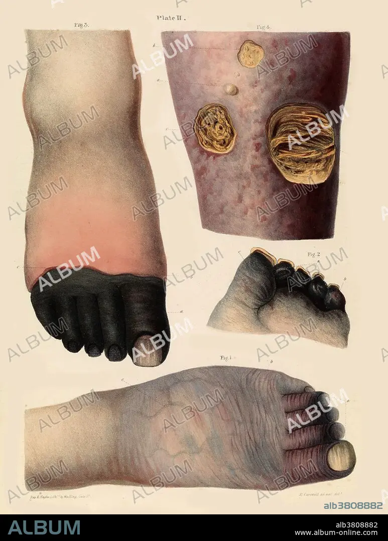
Titre: Gangrene, Illustration, 1830s
Légende: Voir la traduction automatique
Gangrene illustration from the 1830s. Figs 1 and 2 (bottom and centre right) show gangrena senilis - discoloration of the toes at the beginning of the disease. Fig 3 (top left) shows gangrene of the toes. Fig 4 (top right) shows mortification of the skin and subjacent cellular tissue from an obstacle to the return of the venous blood in consequence of disease of the heart. Robert Carswell.
Gangrene illustration from the 1830s. Figs 1 and 2 (bottom and centre right) show gangrena senilis - discoloration of the toes at the beginning of the disease. Fig 3 (top left) shows gangrene of the toes. Fig 4 (top right) shows mortification of the skin and subjacent cellular tissue from an obstacle to the return of the venous blood in consequence of disease of the heart. Robert Carswell.
Crédit: Album / Science Source / Wellcome Images
Autorisations: ? Autorisation de modèle: Non - ? Autorisation de propriété: Non
Questions sur les droits?
Questions sur les droits?
Taille de l'image: 4952 × 6556 px | 92.9 MB
Taille d'impression: 41.9 × 55.5 cm | 1949.6 × 2581.1 in (300 dpi)
Mots clés: CORPS PIEDS • DERMATOLOGIE • DESORDRE • GANGRENE • ILLUSTRATION • JAMBE • JAMBE, ANATOMIE • JAMBES • MALADIE DE PEAU • MEDICAL • ORTEIL • PATHOLOGIE • PERSONNE • PIED • PIEDS
