alb3786576
Breast Anatomy, Illustration
| Partager |
|---|
 Pinterest Pinterest |
 Twitter Twitter |
 Facebook Facebook |
 Copier le lien Copier le lien |
 Email Email |
|
Ajouter à une autre Lightbox |
|
Ajouter à une autre Lightbox |


Avez-vous déjà un compte? S'identifier
Vous n'avez pas de compte ? S'inscrire
Acheter cette image

Titre:
Breast Anatomy, Illustration
Légende:
Voir la traduction automatique
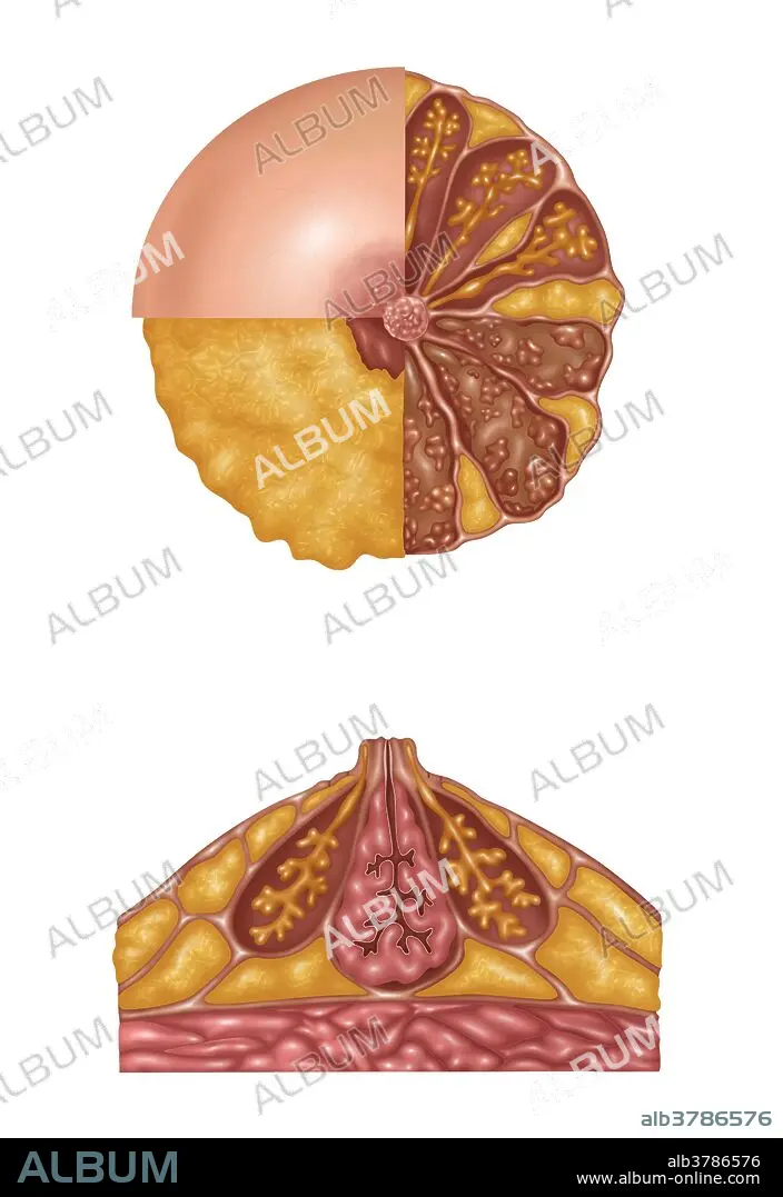
Illustration detailing the anatomy of a female breast from a front view (top) and a side view (bottom). The top image is in three sections. The areola, montgomery's tubercules, and nipple are in the light pink section. The nipple and subareolar musculature and subcutaneous fat are in the orange section. On the right half is: mammary fat (orange), lactiferous ducts (orange stems), acini (end of orange stems), ampulla (on orange stem), coopers ligaments (light pink strands), lobe and lobules (brown outlines and pink groups inside the brown), and interlobular connective tissue (brown area). The bottom image shows montgomery's gland and superficial fascia (outer lining), subcutaneous fat (orange outer sections), ampulla and lactiferous duct and connective tissue (pink center), coopers ligaments, mammary fat (orange bottom sections), pectoral fascia, and pectoralis major (pink at bottom).
Crédit:
Album / Science Source / Gwen Shockey
Autorisations:
Modèle: Non - Propriété: Non
Questions sur les droits?
Questions sur les droits?
Taille de l'image:
4200 x 6108 px | 73.4 MB
Taille d'impression:
35.6 x 51.7 cm | 14.0 x 20.4 in (300 dpi)
Mots clés:
