alb3818128
Nerve Cell from Spinal Cord, Deiters, 1865
| Partager |
|---|
 Pinterest Pinterest |
 Twitter Twitter |
 Facebook Facebook |
 Copier le lien Copier le lien |
 Email Email |
|
Ajouter à une autre Lightbox |
|
Ajouter à une autre Lightbox |


Avez-vous déjà un compte? S'identifier
Vous n'avez pas de compte ? S'inscrire
Acheter cette image

Titre:
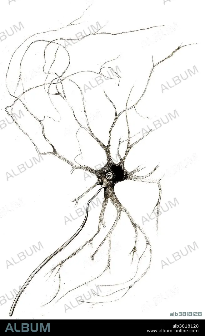
Nerve Cell from Spinal Cord, Deiters, 1865
Légende:
Voir la traduction automatique
Nerve cell from anterior horn of spinal cord grey matter. Drawn by Otto Friedrich Carl Deiters in 1865. Deiters (1834-1863) was a German neuroanatomist, remembered for his microscopic research of the brain and spinal cord. His name is lent to the "nucleus of Deiters," also called the lateral vestibular nucleus, and "Deiters' cells," structures that are associated with outer hair cells in the cochlea of the inner ear.
Crédit:
Album / Science Source / Wellcome Images
Autorisations:
Modèle: Non - Propriété: Non
Questions sur les droits?
Questions sur les droits?
Taille de l'image:
2430 x 3792 px | 26.4 MB
Taille d'impression:
20.6 x 32.1 cm | 8.1 x 12.6 in (300 dpi)
Mots clés:
