alb10657099
Lingual Thyroid, CT Scan
| Partager |
|---|
 Pinterest Pinterest |
 Twitter Twitter |
 Facebook Facebook |
 Copier le lien Copier le lien |
 Email Email |
|
Ajouter à une autre Lightbox |
|
Ajouter à une autre Lightbox |


Avez-vous déjà un compte? S'identifier
Vous n'avez pas de compte ? S'inscrire
Acheter cette image.
Sélectionnez l'usage:

Titre:
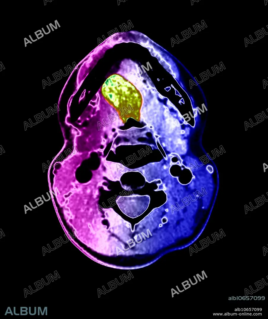
Lingual Thyroid, CT Scan
Légende:
Voir la traduction automatique
This colour-enhanced, axial (cross-sectional) CT image through the oral cavity demonstrates a large, abnormal density (green) in the tongue. This represents lingual thyroid tissue. This patient had no thyroid tissue in the expected region of the lower neck. The thyroid gland develops near the foramen cecum of the tongue and normally descends along a tract called the thyroglossal duct to the lower neck. Rarely, this normal descent does not occur and thyroid tissue remains in the tongue, but remnants of thyroid tissue and the duct itself may occur anywhere along the tract. Often, however, no thyroid tissue is present in the lower neck, only in the tongue base, as in this case.
Personnalités:
Crédit:
Album / Science Source / Living Art Enterprises
Autorisations:
Modèle: Non - Propriété: Non
Questions sur les droits?
Questions sur les droits?
Taille de l'image:
4200 x 4755 px | 57.1 MB
Taille d'impression:
35.6 x 40.3 cm | 14.0 x 15.8 in (300 dpi)
Mots clés:
