alb9203050
Trigeminal Nerve, Illustration
| Partager |
|---|
 Pinterest Pinterest |
 Twitter Twitter |
 Facebook Facebook |
 Copier le lien Copier le lien |
 Email Email |
|
Ajouter à une autre Lightbox |
|
Ajouter à une autre Lightbox |


Avez-vous déjà un compte? S'identifier
Vous n'avez pas de compte ? S'inscrire
Acheter cette image.
Sélectionnez l'usage:

Titre: Trigeminal Nerve, Illustration
Légende: Voir la traduction automatique
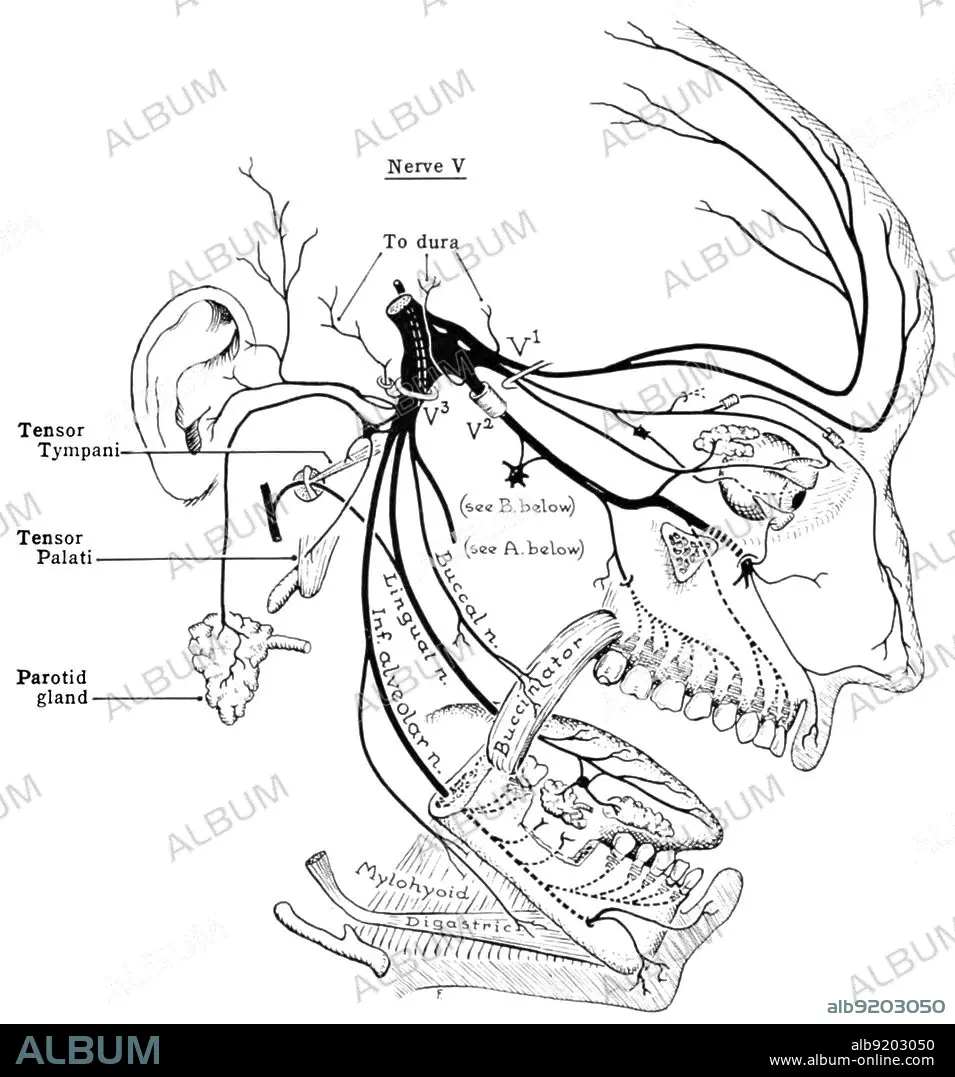
Diagram of the trigeminal nerve. From An Atlas of Anatomy by John Charles Boileu Grant, 1962. The trigeminal nerve (the fifth cranial nerve, or simply CN V) is a nerve responsible for sensation in the face and motor functions such as biting and chewing; it is the largest of the cranial nerves. The three major branches of the trigeminal nerve???the ophthalmic nerve (V1), the maxillary nerve (V2) and the mandibular nerve (V3)???converge on the trigeminal ganglion (also called the semilunar ganglion or gasserian ganglion), located within Meckel's cave and containing the cell bodies of incoming sensory-nerve fibers.
Diagram of the trigeminal nerve. From An Atlas of Anatomy by John Charles Boileu Grant, 1962. The trigeminal nerve (the fifth cranial nerve, or simply CN V) is a nerve responsible for sensation in the face and motor functions such as biting and chewing; it is the largest of the cranial nerves. The three major branches of the trigeminal nerve???the ophthalmic nerve (V1), the maxillary nerve (V2) and the mandibular nerve (V3)???converge on the trigeminal ganglion (also called the semilunar ganglion or gasserian ganglion), located within Meckel's cave and containing the cell bodies of incoming sensory-nerve fibers.
Crédit: Album / Science Source
Autorisations: ? Autorisation de modèle: Non - ? Autorisation de propriété: Non
Questions sur les droits?
Questions sur les droits?
Taille de l'image: 1720 × 1844 px | 9.1 MB
Taille d'impression: 14.6 × 15.6 cm | 677.2 × 726.0 in (300 dpi)
Mots clés: ANATOMIE • DIAGRAMME • HUMAIN • ILLUSTRATION • MAL DE TETE • MIGRAINE • PERSONNE
