alb3773210
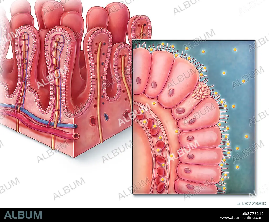
Intestinal Villi, illustration
| Partager |
|---|
 Pinterest Pinterest |
 Twitter Twitter |
 Facebook Facebook |
 Copier le lien Copier le lien |
 Email Email |
|
Ajouter à une autre Lightbox |
|
Ajouter à une autre Lightbox |


Avez-vous déjà un compte? S'identifier
Vous n'avez pas de compte ? S'inscrire
Acheter cette image.
Sélectionnez l'usage:

Titre: Intestinal Villi, illustration
Légende: Voir la traduction automatique
An illustrated section of villi from the small intestine as well as a close up view of a single villus. Villi are finger-like projections that extend into the lumen of the small intestine, increasing surface area for greater nutrient absorption. Each villus is lined with columnar epithelium known as enterocytes, with each cell containing microvilli to further increase surface area. Digested nutrients are absorbed into nearby capillaries so that it can then be transported to the rest of the body.
An illustrated section of villi from the small intestine as well as a close up view of a single villus. Villi are finger-like projections that extend into the lumen of the small intestine, increasing surface area for greater nutrient absorption. Each villus is lined with columnar epithelium known as enterocytes, with each cell containing microvilli to further increase surface area. Digested nutrients are absorbed into nearby capillaries so that it can then be transported to the rest of the body.
Crédit: Album / Science Source / Evan Oto
Autorisations: ? Autorisation de modèle: Non - ? Autorisation de propriété: Non
Questions sur les droits?
Questions sur les droits?
Taille de l'image: 3300 × 2550 px | 24.1 MB
Taille d'impression: 27.9 × 21.6 cm | 1299.2 × 1003.9 in (300 dpi)
