alb10652681
Abdominal aorta, calcification, CT angiography
| Partager |
|---|
 Pinterest Pinterest |
 Twitter Twitter |
 Facebook Facebook |
 Copier le lien Copier le lien |
 Email Email |
|
Ajouter à une autre Lightbox |
|
Ajouter à une autre Lightbox |


Avez-vous déjà un compte? S'identifier
Vous n'avez pas de compte ? S'inscrire
Acheter cette image.
Sélectionnez l'usage:

Titre: Abdominal aorta, calcification, CT angiography
Légende: Voir la traduction automatique
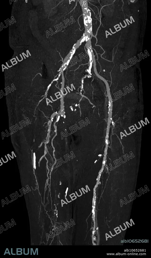
CT angiography of the lower extremities reveals dense calcification (bright white plaques) of abdominal aorta, femoral and popliteal arteries in a 68 year old dialysis patient with chronic renal failure and secondary hyperparathyroidism. The right femoral and popliteal arteries are occluded and collateral vessels are visible in the distal thigh.
CT angiography of the lower extremities reveals dense calcification (bright white plaques) of abdominal aorta, femoral and popliteal arteries in a 68 year old dialysis patient with chronic renal failure and secondary hyperparathyroidism. The right femoral and popliteal arteries are occluded and collateral vessels are visible in the distal thigh.
Crédit: Album / Science Source / Steven Needell
Autorisations: ? Autorisation de modèle: Non - ? Autorisation de propriété: Non
Questions sur les droits?
Questions sur les droits?
Taille de l'image: 2337 × 3787 px | 25.3 MB
Taille d'impression: 19.8 × 32.1 cm | 920.1 × 1490.9 in (300 dpi)
